 Home /
 At Retina Institute of Virginia, we understand how important clear vision is for your quality of life. Our dedicated team of experienced eye doctors is here to guide you through every step of your retina treatment journey.


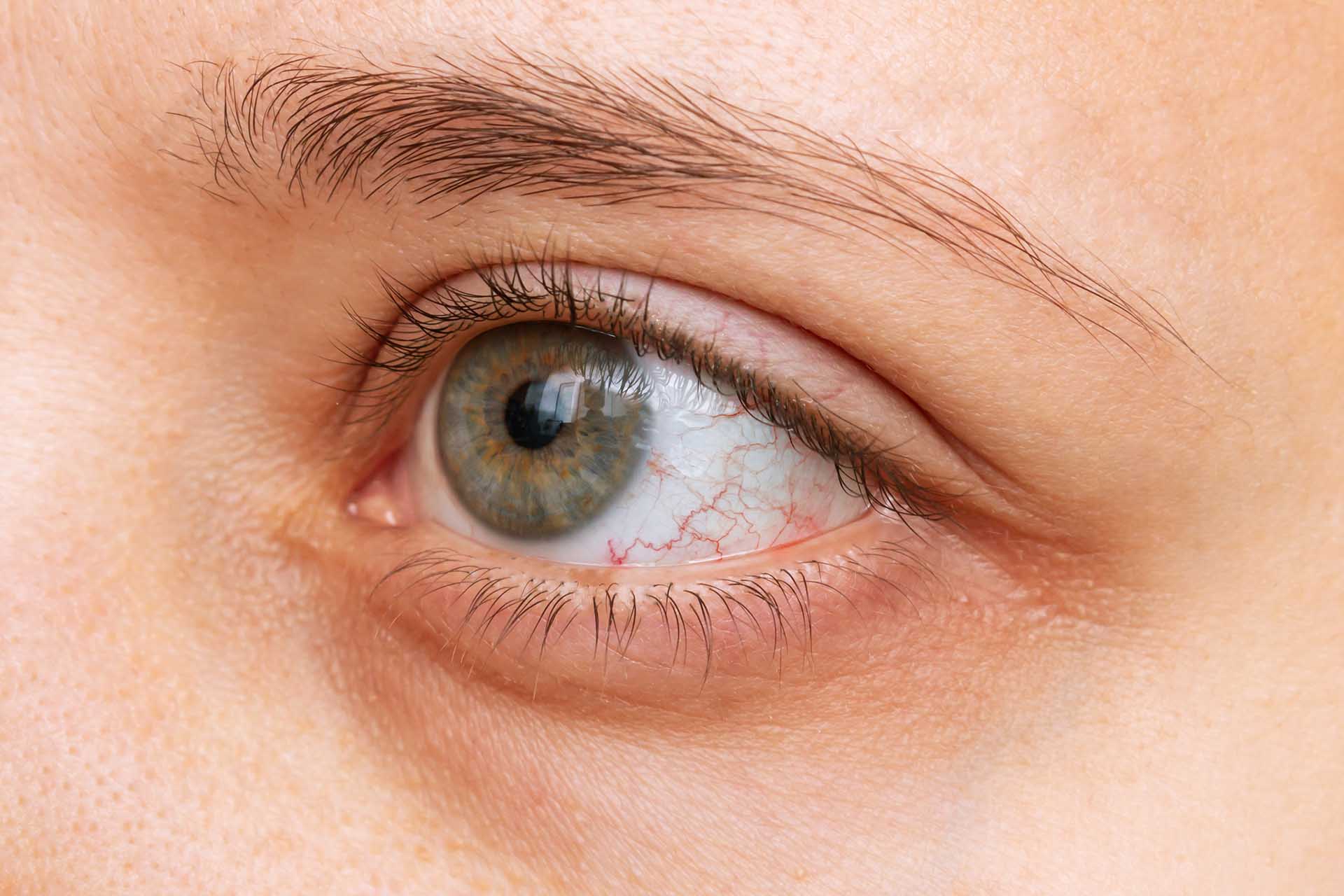
A thin layer of tissue called the retina covers the back inside wall of the eye. When the cornea and lens at the front of the eye focus light on the retina, a picture is taken and sent to the brain through the optic nerve to be interpreted. Thus, the retina is the “seeing tissue” of the eye.
The retina has two parts: the peripheral retina and the macula.
The macula is the center of the retina, while the large area surrounding the macula is the peripheral retina. The peripheral retina allows us to see out of the corner of the eye but is unable to see detail clearly. We cannot drive, read or see facial features with the peripheral retina.
Although small, the macula is one hundred times more sensitive to detail than the peripheral retina. The macula is required to see fine detail and it must be healthy to work properly.

The vitreous is a jelly-like substance that fills the cavity between the lens and the back of the eye. Also called the vitreous humor. As we age, the vitreous ages along with the rest of the body, and these aging changes can sometimes lead to problems with vision and even damage to the retina. That is why we often speak of patients with vitreo-retinal disease.”
Age-related macular degeneration (AMD) is a common eye condition and a leading cause of vision loss among older adults. It results from degeneration of the macular tissue and can cause central vision loss, blind spots, changes in color perception, and distorted vision. There are two general categories of AMD, the “wet” or exudative kind, and the “dry” or atrophic kind. The “wet” form of the disease is associated with abnormal blood vessel growth that often leads to severe vision loss. It is often treated with ocular injections. The “dry” form of the disease is treated with careful observation and vitamin therapy. Dry AMD can sometimes turn into wet AMD, and when that happens, prompt diagnosis and therapy are essential to maximize visual outcomes.
Diabetes is a chronic condition that affects the body’s ability to process blood sugar. People with diabetes are at risk for diabetic retinopathy, an eye disease that damages the smallest retinal blood vessels. Diabetic retinopathy has several distinct stages with varying symptoms. Early detection is important to prevent permanent vision loss. Learn more about diabetic retinopathy and available treatment options at Retina Institute of Virginia on our Diabetic Eye Disease page.
Endophthalmitis is an infection of the eye and is also an ocular emergency. Infections can spread to the eye from infections elsewhere in the body, can occur because of trauma to the eye, and can also be a complication of ocular injections or eye surgery. The prognosis of untreated endophthalmitis is often very poor, with a high likelihood of loss of vision, loss of the eye itself, and even the spread of infection from the eye to other parts of the body. The diagnosis of infectious endophthalmitis involves careful examination of the eye which often leads to a “tap and inject” procedure where a small amount of fluid is removed from the eye for evaluation in a laboratory to identify the microbe that is causing the infection.
Flashes and floaters are common visual disturbances that appear as flashes of light or as dark specks or strands. Flashes and floaters are typically the result of changes in the vitreous gel but can also be signs of more serious disease. They are often harmless, but it is important to have them evaluated by an experienced eye doctor with expertise in vitreoretinal disease.
The macula is delicate, and if it becomes swollen or inflamed, a condition called macular edema can occur. This condition causes decreased central vision and may also cause redness and discomfort. Macular edema is often associated with other retinal diseases and can be treated with eye drops, injectable medications, and sometimes surgery.
A macular hole occurs when the vitreous pulls away from the retina in a way that causes a gap to form in the macula. A macular hole causes blurriness in central vision, makes straight lines look wavy, and creates difficulty reading and performing daily tasks. Surgery may be used to repair a macular hole.
Shrinking of the vitreous is a natural part of the aging process and can lead to the development of scar tissue that may cause the macula to wrinkle or pucker. This condition typically causes symptoms such as mild blurriness or distortion in vision. If a macular pucker does not resolve on its own and it causes significant symptoms, it can be treated with surgery.

A retinal tear can occur due to aging or an eye injury. Similar to retinal detachment, a tear in the retina can cause shadows in vision or flashes. If left untreated, a retinal tear can lead to retinal detachment. Retinal tears often require treatment–but not always.
A detached retina occurs when the retina separates from the underlying layers of the eye, which can cause symptoms including vision loss, sudden shadows, bright flashes, and floaters. Retinal detachment can happen gradually or suddenly and can be associated with inflammatory conditions, vitreous aging changes, or an eye injury. Retinal detachment is a medical emergency and should be diagnosed immediately. A variety of surgical techniques can be employed to treat retinal detachment and retinal specialists will discuss and offer the best alternatives suited to your specific details. The timing of retinal detachment repair can range from emergent (within a day or two) to within many weeks or even months.
The small blood vessels of the retina transfer nutrients and oxygen to the retina, and are essential for proper retinal function. Retinal arteries supply blood to the retina and retinal veins carry it away. The blood vessels of the retina branch out from the larger blood vessels that provide blood supply to the rest of the brain.
If a retinal artery becomes blocked, it is called a retinal artery occlusion (RAO). RAO is a medical emergency and can be thought of as a stroke of the retina. Partial or complete visual loss may become permanent without immediate diagnosis and treatment. RAO patients also have a high risk for strokes to other parts of the brain. The eye doctors often coordinate care with other medical doctors to give RAO patients the best possible chances to recover or preserve vision.
If a retinal vein becomes blocked, it causes retinal vein occlusion, or RVO. RVO is sometimes associated with other ocular or medical conditions, and preventative care for risk factors such as high blood pressure, diabetes, glaucoma, macular edema, or inflammatory conditions is an important part of caring for patients with RVO. The treatment for RVO depends on the type, location, and severity of the blockage in the retinal vein and may consist of observation, injections of medication, laser surgery and sometimes microsurgery.
Uveitis is a disease where the eye or a portion of the eye becomes inflamed. When the inflammation spreads to the vitreous and retina, the patient is said to have posterior uveitis, which may represent a more serious disease. Uveitis can be caused by infection, autoimmune disease, or trauma or may not have any identifiable cause. Even after extensive medical and laboratory testing, about half of all uveitis patients never have the cause of their uveitis identified.

Retinitis pigmentosa is an uncommon, inherited disease where the retina gradually degenerates. The retina is the light-sensing cells that line the back of your eye and converts light rays into signals that are transmitted through the optic nerve to the brain, where they are recognized as images.
Retinitis pigmentosa is a slow and progressive disease, which causes low vision. Some symptoms include:
Every person will experience vision change at a different pace depending on the genetic makeup of the disorder.